- Shop A054 / 147-189 Brisbane Road. Biggera Waters, QLD 4216
- admin@htdoctors.com.au
- Contact: 07 5222 7117

Australia have the highest melanoma rates in the world, and Queensland consistently leads the nation in diagnoses. On the Gold Coast, where outdoor living and high UV exposure are part of daily life, skin cancer is not a distant risk. It is something that affects one in every two Australians before the age of 70. The vast majority of skin cancer deaths are preventable with early detection, yet thousands of people are diagnosed each year with cancers that had been present and growing unnoticed for months or even years. At Harbour Town Doctors, our skin cancer clinic provides thorough professional checks for patients across the Gold Coast. Here are the warning signs that are most commonly missed, and what you need to know to protect yourself and your family.
Most Australians are aware that skin cancer exists and that sun protection matters. Fewer are aware that skin cancer can look nothing like what they expect, that it can appear in places they never think to check, or that several of the most dangerous forms develop with no symptoms at all until they are advanced.
According to Cancer Australia, melanoma is the third most commonly diagnosed cancer in Australia and it is estimated to cause around 1,455 deaths in 2025. Queensland has consistently recorded Australia’s highest melanoma incidence rate, at approximately 64 cases per 100,000 people compared to a national average of around 48. Living on the Gold Coast puts you directly in one of the highest risk regions in the country.
The problem is not only UV exposure. It is also recognition. People delay seeking medical attention because a spot does not look the way they expect skin cancer to look. The following warning signs are those most frequently overlooked.
Many Australians associate skin cancer with a dark, irregular mole. This is an understandable but dangerous oversimplification. The three most common forms of skin cancer look very different from each other, and two of them are rarely darkly pigmented at all.
Basal cell carcinoma (BCC) is the most common skin cancer in Australia. It typically appears as a pale, pearly, or pink spot or lump, sometimes with a translucent or shiny surface. It may look like a small scar or a patch of skin that seems slightly unusual. It rarely causes pain. People often dismiss it as a minor blemish, a cyst, or simply a part of ageing skin. BCCs grow slowly and very rarely spread to other parts of the body, but if left untreated they can cause significant local tissue destruction.
Squamous cell carcinoma (SCC) often appears as a firm pink or red lump, a rough scaly patch, a wart like growth, or an open sore that fails to heal completely. SCCs grow faster than BCCs and carry a greater risk of spreading if not treated. They are commonly found on areas of chronic sun damage including the face, ears, scalp, neck, and backs of the hands.
Neither BCC nor SCC looks like the classic dark melanoma that most people picture. Learning to recognise these less obvious presentations is essential.
A wound, ulcer, or sore that opens, crusts over, and then reopens without fully healing is one of the most consistently missed warning signs of skin cancer. It is frequently attributed to an insect bite, a minor skin injury, or a reaction to a product, and weeks pass before a doctor is consulted.
Both SCC and BCC can present as a non healing sore. In some cases, the lesion bleeds easily when scratched or rubbed and then appears to be healing, only to open again. Any sore or ulcer on the skin that has not healed within three to four weeks should be assessed by a GP. If you have a lesion that is doing this, our skin cancer clinic at Harbour Town Doctors can assess it promptly with dermoscopic examination.
The ABCDE criteria were developed to help people and doctors identify suspicious changes in moles and pigmented lesions. Each letter identifies a specific feature that warrants further assessment.
A normal mole, if you drew a line through the centre, should produce two roughly equal halves. An asymmetric mole, where one half does not match the other, is a warning sign. The asymmetry may be subtle and is easy to miss if you are not specifically looking for it.
Normal moles have smooth, clearly defined edges. A mole with an irregular, ragged, notched, or blurred border is cause for concern. The edges may appear to fade into the surrounding skin in an uneven way or have a scalloped appearance.
A benign mole is usually a single shade of brown or tan. Multiple colours within a single lesion, including varying shades of brown, black, red, white, or blue, indicate that different cell populations may be present. Uneven colour distribution within the same spot is a significant warning sign.
Melanomas are often larger than 6 millimetres in diameter when diagnosed, roughly the size of a pencil eraser. However, some melanomas are smaller when detected, and this criterion should not be used in isolation. Any lesion that appears to be growing in size warrants attention regardless of its current measurement.
This is arguably the most important criterion of all. A lesion that is changing in size, shape, colour, or texture, or that begins to bleed, itch, crust, or feel different, should be seen by a doctor regardless of how it scores against the other criteria. Change is the key signal. Normal moles remain stable over years. An evolving lesion demands prompt assessment.
The ABCDE system is a powerful guide but is not exhaustive. Some melanomas, particularly nodular melanomas, can develop rapidly without following the typical ABCDE pattern. Any new or changing lesion that concerns you should be assessed professionally.
Nodular melanoma is the second most common subtype of melanoma but accounts for a disproportionate number of melanoma deaths because it behaves differently from the standard ABCDE pattern. Rather than spreading outward across the skin surface, nodular melanoma grows downward rapidly. As described by Cancer Council Australia, it typically presents as a raised, dome shaped, firm nodule that may be black, dark brown, blue, or even pink, red, or flesh coloured. It can reach a dangerous thickness within weeks to months.
The danger of nodular melanoma is that it may appear relatively small and recently formed when it is already deep. Because it does not spread laterally across the surface first, it does not develop the irregular borders and colour variations that the ABCDE guide is designed to detect. Many patients describe it as a new lump that appeared quickly and seemed to be enlarging. If you have any rapidly growing raised lesion on your skin, see a doctor urgently.
Most people check their face, arms, and back. Far fewer check their scalp, the spaces between their toes, the soles of their feet, the palms of their hands, under their fingernails and toenails, the skin of the lower legs, or the areas of the scalp covered by hair. All of these sites are known locations for melanoma development.
Acral lentiginous melanoma is a subtype that specifically affects the palms, soles, and nail beds. It accounts for a small percentage of all melanomas in Australia but is particularly dangerous because it is so frequently discovered late. A dark streak running lengthwise under a fingernail or toenail that is not explained by recent injury should be assessed by a doctor.
Scalp melanomas are also commonly missed because they are concealed by hair. Use a comb to part the hair systematically when doing a self check, or ask a partner to help examine areas you cannot easily see yourself. Your GP will check your scalp during a full skin cancer examination.
Amelanotic melanoma is a form of melanoma that lacks the dark pigmentation most people associate with the disease. It can appear pink, red, flesh coloured, or only faintly pigmented. Because it looks nothing like a traditional mole or dark lesion, it is frequently mistaken for a pimple, a minor skin infection, a pink scar, or even an inflammatory patch.
Lentigo maligna melanoma is another subtype that is frequently missed. It develops slowly over years within a flat, irregularly shaped, unevenly pigmented patch on sun damaged skin, most commonly the face or neck of older adults. It may resemble an age spot or a sun freckle for a long time before becoming invasive.
The common thread in all missed diagnoses is the assumption that a lesion that looks familiar or benign does not need to be checked. If a spot is new, if it is changing, or if it simply does not look quite right to you, have it seen.
Skin cancer is not usually painful in its early stages. However, some lesions do itch, particularly BCCs and superficial spreading melanomas. A spot that bleeds with minimal contact, such as when towelling dry or rolling over in bed at night, is a notable warning sign that is easy to attribute to friction or dry skin.
A lesion that crusts and appears to heal slightly before reforming a crust, or one that feels raised, firm, or different in texture from surrounding skin, should be added to the list of lesions to discuss with your doctor at your next visit, or sooner if the change seems rapid.
While anyone can develop skin cancer, certain factors significantly elevate the risk. On the Gold Coast, where UV levels regularly reach extreme ratings and outdoor activity is year round, the following groups should be particularly vigilant.
A self examination every three months is strongly recommended and can help you identify changes between professional checks. However, it has clear limitations. Most people cannot easily view their entire scalp, back, or the backs of their legs. Self assessment also lacks the magnification and specialised lighting that dermoscopy provides.
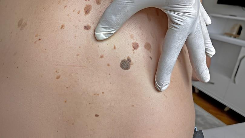
A trained doctor using a dermoscope can identify features within a lesion that are completely invisible to the naked eye, including vascular patterns, regression structures, and pigment network irregularities that are strongly associated with malignancy. Professional conduct comprehensive, full body examinations using dermoscopy and provide clear documentation of any lesions identified, along with recommendations for monitoring or biopsy where appropriate.
Do not wait and watch to see if a suspicious spot gets worse. Do not apply creams or treat it yourself. Do not assume that because it does not hurt it is not serious. See a doctor.
The earlier skin cancer is diagnosed, the simpler and more effective the treatment. Melanoma that is confined to the outermost layer of skin has a five year survival rate approaching 100 percent. Once melanoma spreads to the lymph nodes or distant organs, outcomes become significantly worse. Speed matters.
At Harbour Town Doctors, general practice and family medicine team can address any skin concerns you have, and bulk billing is available for eligible patients.
Skin cancer doesn’t always show obvious warning signs. It can appear as a scar, a pimple, an age spot, or a slow-healing wound. With high UV exposure on the Gold Coast, regular skin checks are essential, not optional. They are one of the smartest and potentially life-saving health decisions you can make each year. contact us today, if you haven’t had a check in the past 12 months or notice any changes.
At least once a year for most adults. Queensland has the highest melanoma rate in Australia. People with fair skin, many moles, or a history of significant sun exposure should consider checks every six months. Do a self check every three months between visits and see your GP promptly for any new or changing spots.
Yes. Acral lentiginous melanoma develops on the palms, soles, and under the nails. Mucosal melanoma can occur in the mouth or genitals. Ocular melanoma forms inside the eye. A thorough skin check covers all body surfaces, not only sun exposed areas.
A normal mole is round or oval, has a smooth and defined edge, is a uniform shade, is smaller than 6 millimetres, and stays stable over time. A melanoma may be asymmetric, have an irregular border, show multiple colours, be larger than 6 millimetres, or be evolving. It can also appear as a completely new spot. When in doubt, get it checked.
Yes. Basal cell carcinoma, the most common skin cancer in Australia, often appears as a pale, pearly, or pink spot that may look shiny or scar like. Squamous cell carcinoma may appear as a firm pink or red lump, a rough scaly patch, or a non healing sore. Any new persistent spot or lump that does not resolve within a few weeks should be assessed by a doctor.
A doctor examines your skin from head to toe, using a dermoscope to inspect lesions in magnified detail. The check takes 15 to 30 minutes and is painless. Suspicious spots may be photographed for monitoring or referred for biopsy. At Harbour Town Doctors, our skin cancer clinic provides thorough checks for patients of all ages on the Gold Coast.
At Harbour Town Doctors, we have been providing comprehensive medical care to patients, utilising a team of experienced doctors and allied healthcare professionals through a continuous envelope of personalised care.
Medical Centre Near Me
24 Hour Medical Centre Near Me
Bulk Billing Medical Centre Near Me
After Hours GP Near Me
GP Near Me
Best Doctors Gold Coast
Doctors Near Me
Bulk Billing Doctor Near Me
Bulk Bill Doctors Near Me
Female Doctors Near Me
Sexual Health Clinic Near Me
Women’s Health Clinic Near Me
Cosmetic Fillers Near Me